
10 Great Reasons to Upgrade to
BIOQUANT LIFE SCIENCE 2023
Is your lab using an earlier version of BIOQUANT? Need to justify an upgrade to the latest version? Here are 10 reasons to upgrade to the latest version of BIOQUANT Life Science.
Reason #1: Confocal and Lightsheet Data
5D NAVIGATOR FOR CONFOCAL AND LIGHTSHEET IMAGE DATA

Multi-channel Confocal Stack of Cortical Bone Innervation by Erica Scheller and Washington University in St. Louis is licensed under CC BY 4.0. To view a copy of this license, visit creativecommons.org.
BIOQUANT is pleased to announce support for a new dimension in musculoskeletal research. BIOQUANT Life Science now supports multi-dimensional image data from confocal and lightsheet microscopes.
Simultaneously, a new generation of microscope systems called Lightsheet microscopes are starting to collect enormous image data sets of entire bones with multiple fluorescent labels and multiple Z depths at sub-micron resolutions.
Reason #2: Better Imaging
Spectacular new cameras
The flagship Prokyon model offers a special 20 megapixel mode through pixel-shifting in addition to the native live image at 1920x1200 60 fps with fanless cooling. The Kapella camera keeps the fanless cooling but saves money by removing the 20 megapixel imaging mode. The Subra model is even more economical by removing the fanless cooling.
TRADE-IN DISCOUNT
BIOQUANT and Jenoptik offer a trade-in discount toward the purchase of a new Jenoptik camera. Every digital camera compatible with a BIOQUANT system qualifies. Contact us for specifics. Discounts vary by camera model.

One Step Brightfield Setup

Configure your Jenoptik camera for brightfield imaging with one click. A new automatic brighfield setup algorithm makes it wonderfully simple to properly adjust your camera for accurate imaging.
Live Image Background Correction

Live uncorrected image. Note that center is slightly brighter than edges. Note fuzzy dots.

Live corrected image. Even lighting across the field of view. No dots.
Cleaner Backgrounds in Fluorescence Imaging

Fluorescence Black Balance parameter set to 0. Spillover fluorescence visible in other parts of the image. Makes it harder to see specific label.

Fluorescence Black Balance set to 25. Dim background fluorescence suppressed. This makes it easier to know which labeled surfaces to measure.
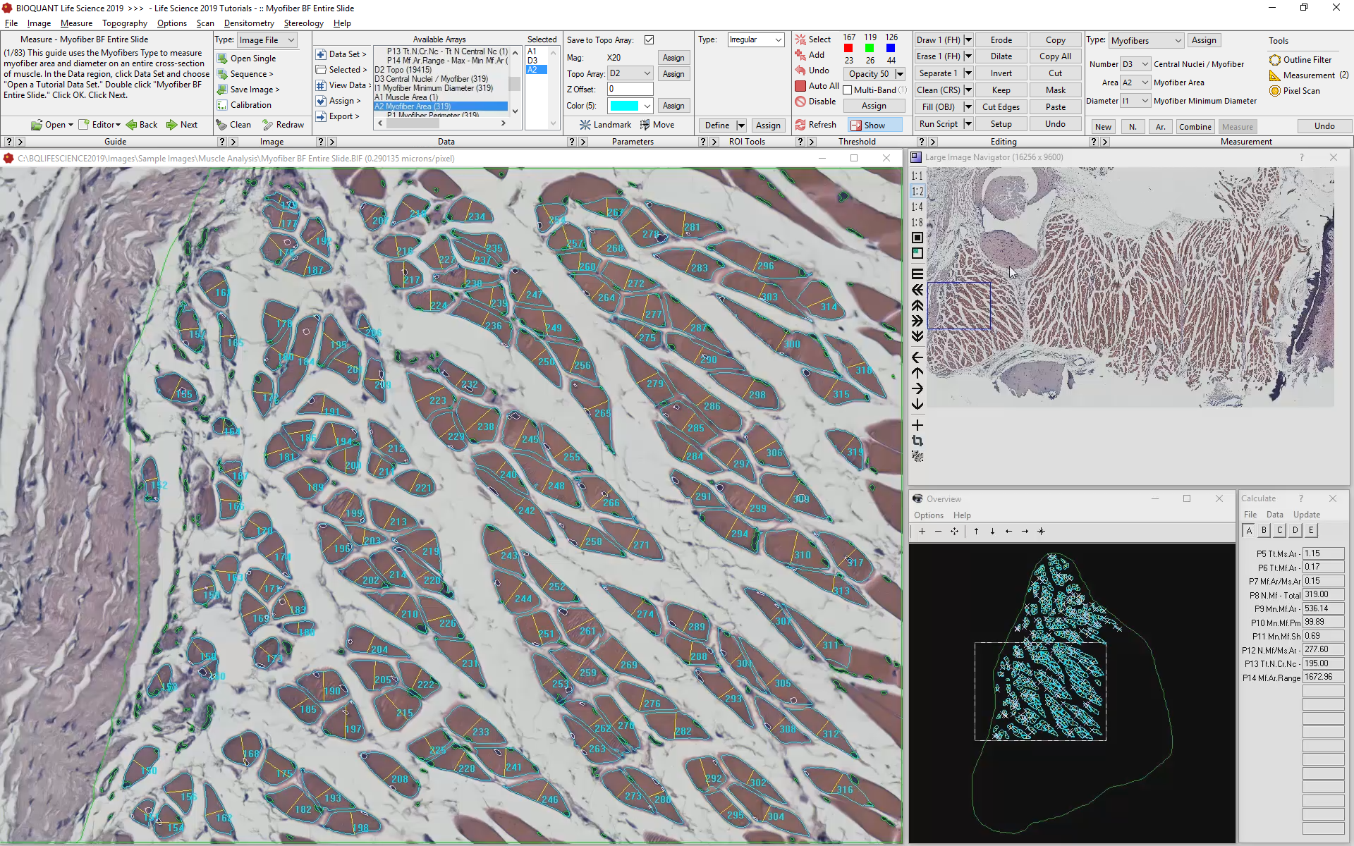
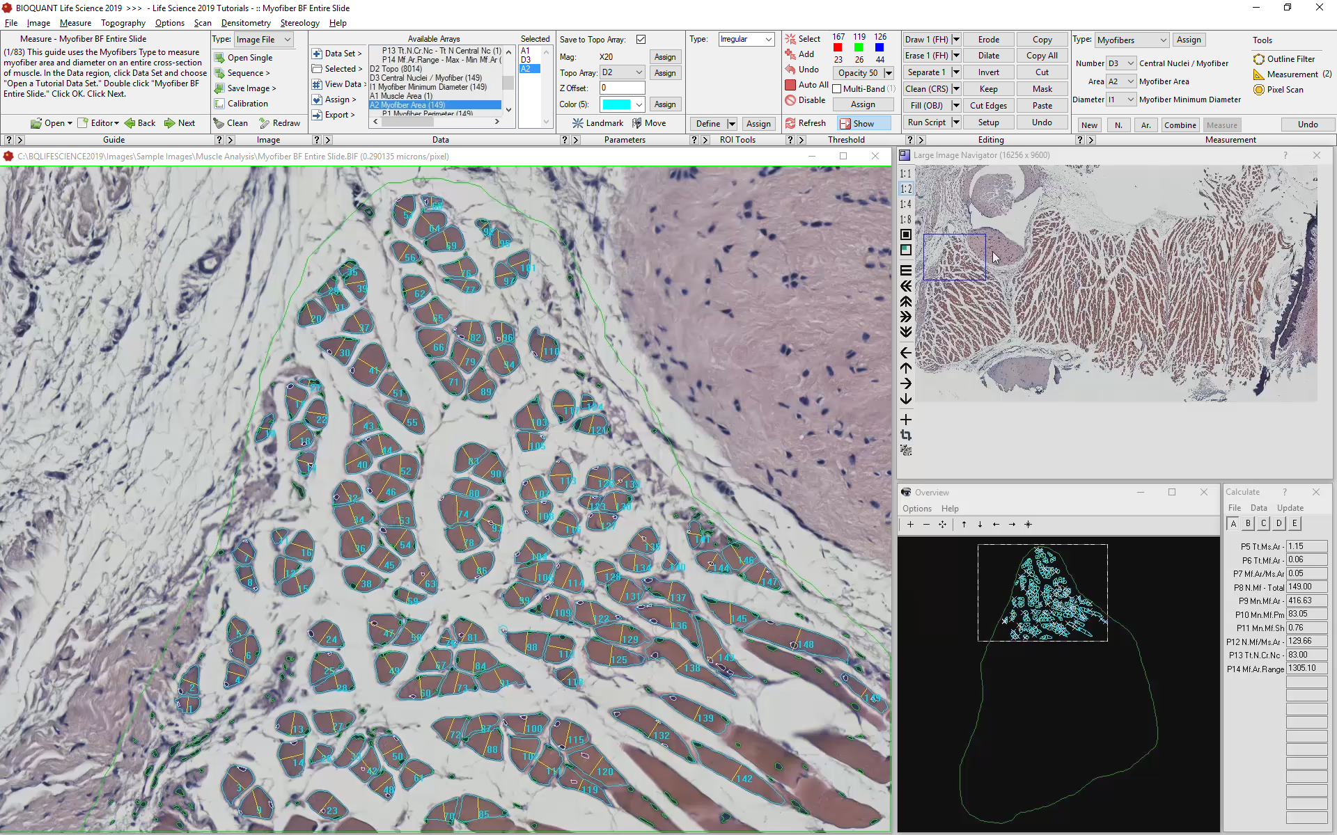
Reason #3: Faster Skeletal Muscle Analysis

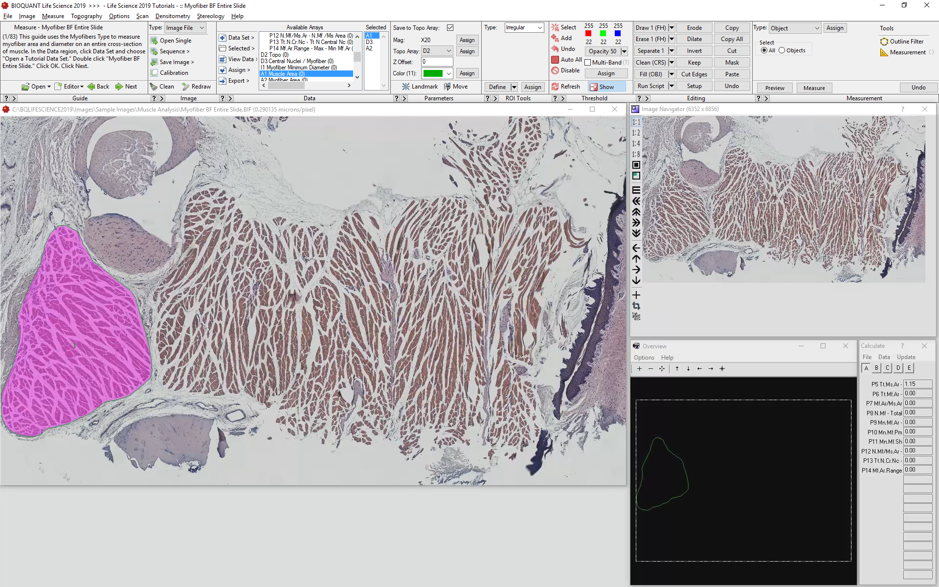
Automated analysis of myofibers depends on a high-contrast label applied to the boundary of each myofiber. Generally this requires a label specific to dystrophin or laminin.
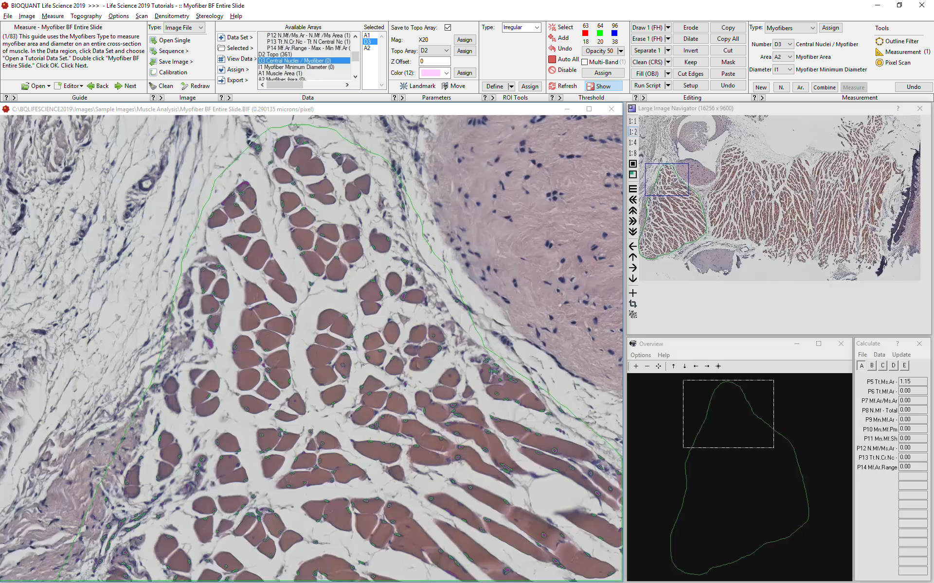
Automatic color thresholding uses this boundary stain to identify the myofibers. Manual editing with a brush and eraser makes it simple to correct mistakes.
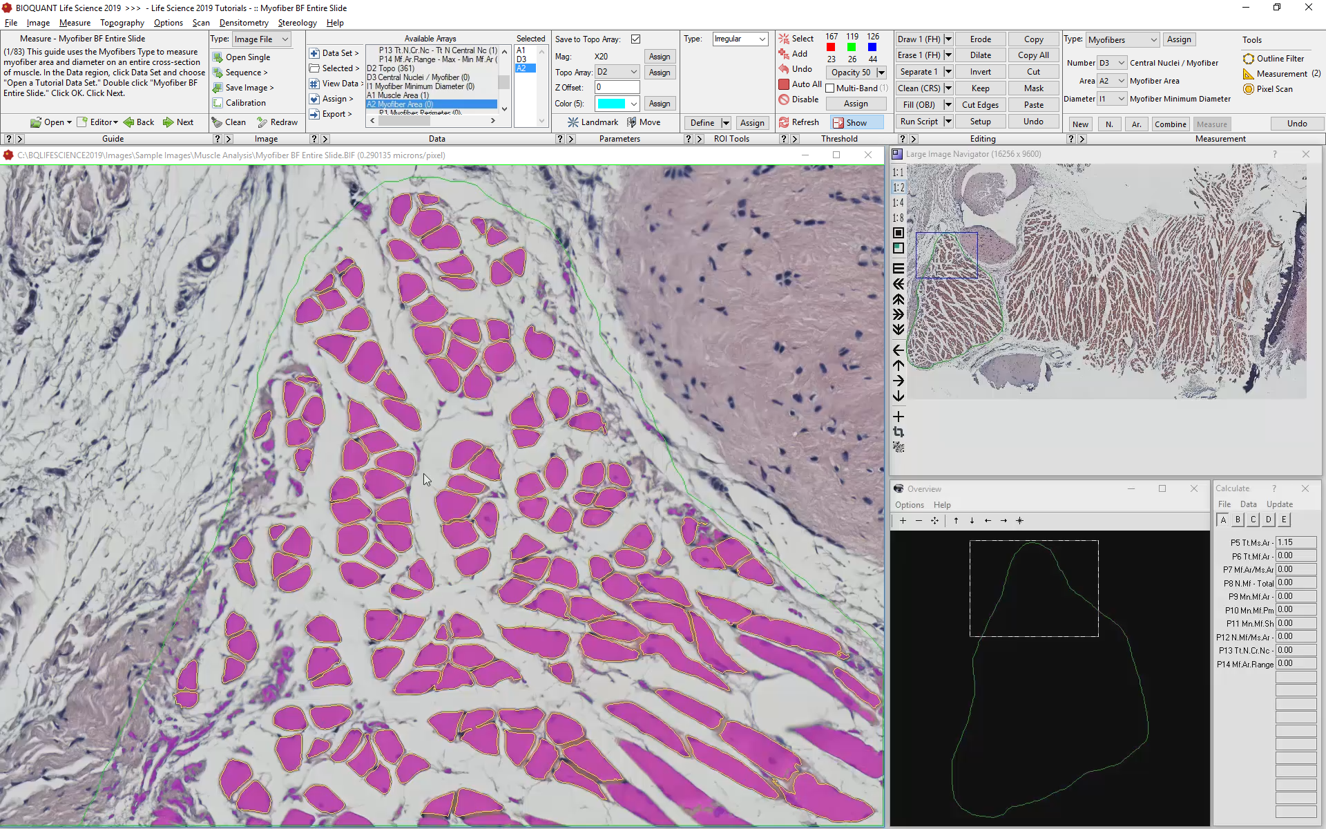
Intelligent filters remove previously measured myofibers and myofibers that are not entirely visible within the field of view.
BIOQUANT simultaneously collects the following data from each myofiber:
Myofiber Cross-sectional Area
Myofiber Shortest Diameter
Myofiber Perimeter
Myofiber Location
Myofiber Circularity
Myofiber Fibertype
Number of Myofibers
Number of Central Nuclei per Myofiber
Reason #4: Faster Thresholding
An accurate threshold is the key to automated measurement. BIOQUANT has made significant progress in automating the creation of accurate thresholds.
Scroll Wheel threshold Adjustment
Fine-tune the Threshold By Scrolling the Mouse Wheel
Starting with the initial color, scrolling the mouse wheel up will search the image for similar colors and add them to the threshold automatically. Scrolling the mouse wheel down will find the colors in the threshold that are least similar to the initial color and remove them automatically.
RAPID THRESHOLD EDITING
Automatically Remove Threshold Associated with Non-specific Staining
The "Clean" button searches the image for all "small" bits of threshold and deletes them. This makes it quick to isolate the important parts of an image from the surrounding tissue despite non-specific background staining.
Keep Just What you Want
The "Keep" button allows you to mark just the parts of the threshold you need and then deletes the rest.
Precise Threshold Editing
Precise multi-band thresholding means you can combine multiple highly specific color ranges into one composite threshold. You can capture all the subtle variations in a stain. You can identify both lightly stained and darkly stained cells at the same time. You can identify cells stained with different fluorochromes at the same time.
Assign Contrasting Threshold Colors
Threshold colors can be assigned to specific arrays now. This means your threshold color will always contrast well with the both the specific stain you're trying to measure and the counterstain.

BIOQUANT Life Science uses a new palette of high-contrast colors.
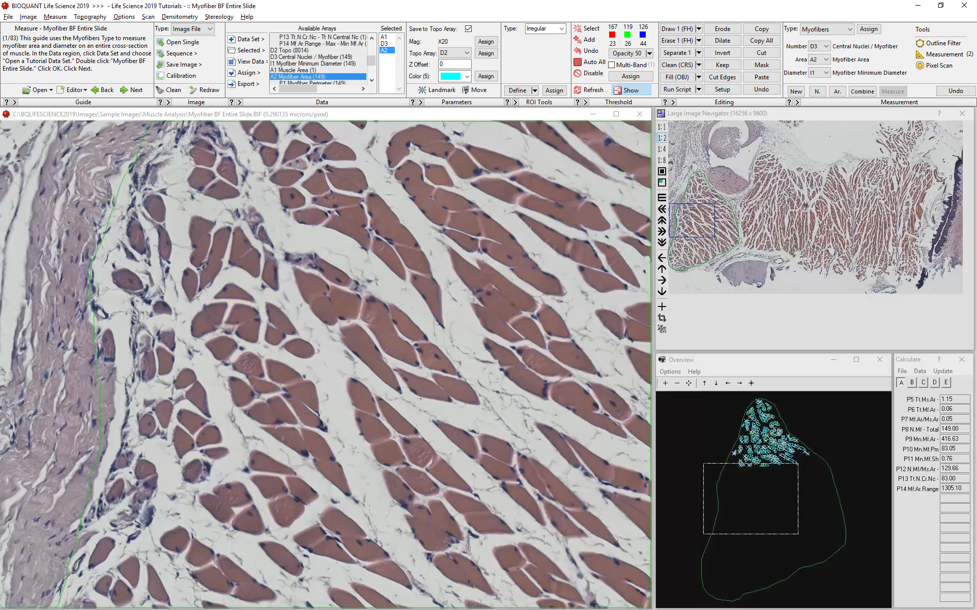
Reason #5: Faster Imaging
4GB Support for digital scans in TIF, BMP, & BIF Format
Open scans in the TIF, BMP, or BIF image formats. This makes it easy to work with digital slides from BIOQUANT SCAN and many other digital pathology scanners. With a 20X lens, that's more than 300 mm2 of tissue.
Crop within the Large Image Navigator
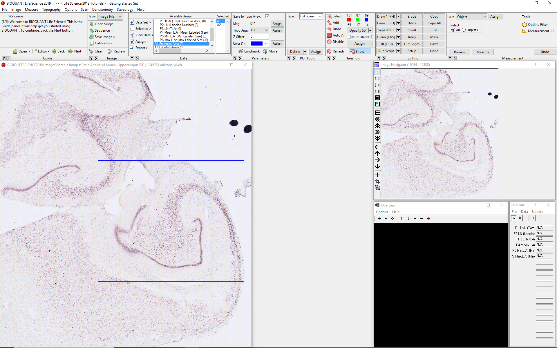
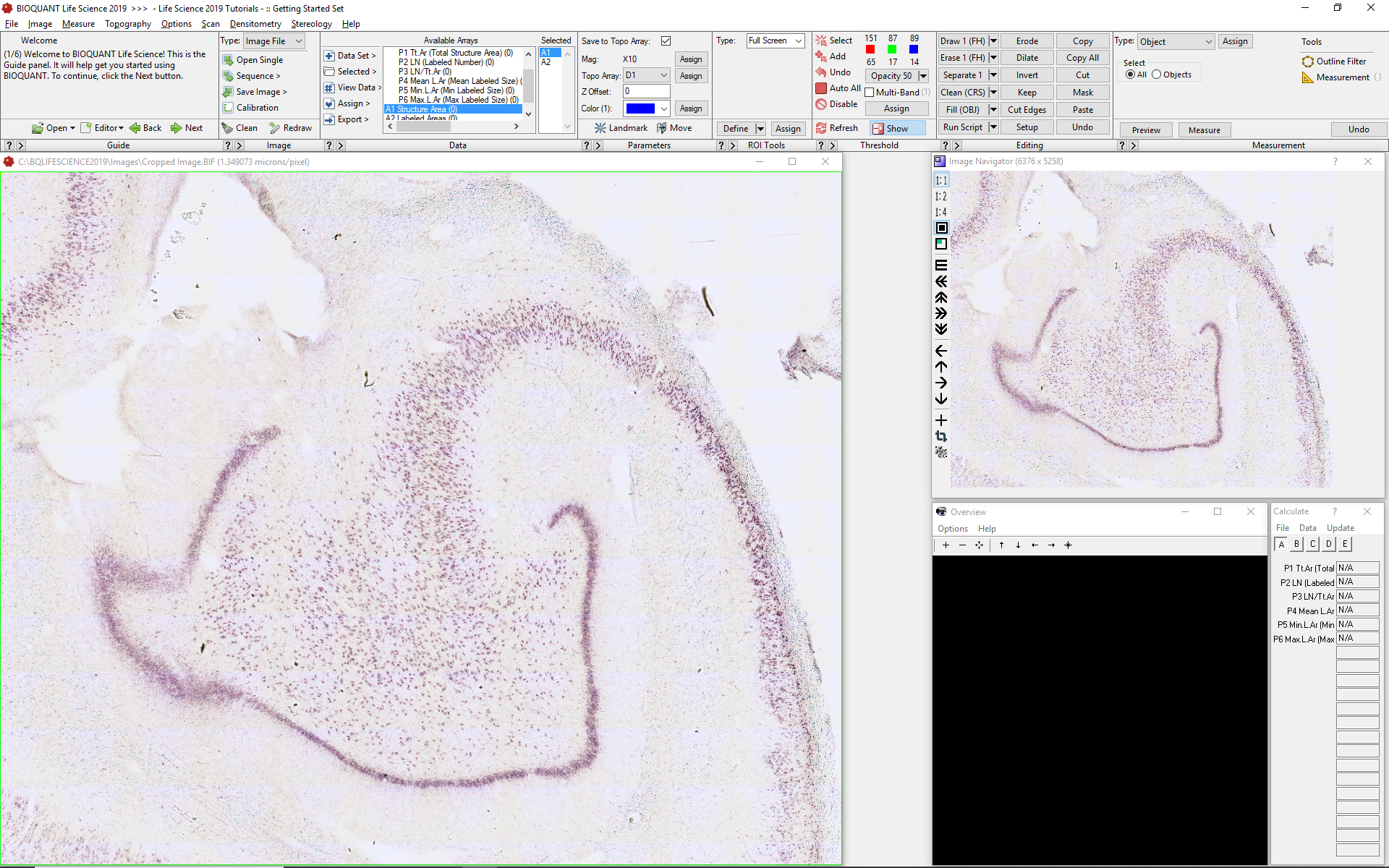
Having access to the entire section, means BIOQUANT can zoom out to rapidly measure large structures hippocampus area, then zoom in to measure cells within any structure. All this combines to make data collection fast.
Store camera Settings in Camera Presets
Camera presets make is simple to switch between brightfield, polarized light, and fluorescence imaging.
Presets store exposure time, gain, and white balance settings to standardize imaging and reduce setup times.
BIOQUANT SCAN - Add features to BIOQUANT Life Science
BIOQUANT SCAN controls a motorized stage with the help of an autofocus camera to automatically scan histology slides or well plate cultures. Analysis of digital scans is faster to perform and easier to train.
BIOQUANT can zoom out to measure referent data at one time for the entire section. Moving around a high magnification is faster and simpler when all you need to do is drag a box around the Large Image Navigator.
Reason #6: Faster Training with Guides and Videos
Learn from Integrated Video Tutorials
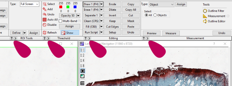
Play Button for Integrated Video Tutorials make it easy to find the help you need when you need it. For example, click the play button in the Measurement region to learn about the various measurement tools. Click the play button in the Threshold region to learn how to automatically identify stained cells.
Get Started quickly with Tutorial Guides

A Warm Welcome
When BIOUQANT starts, a welcome screen appears. This allows students to start one of the many tutorials, continue where they left off, or start a new project.
Start a Tutorial
BIOQUANT comes with an ever-expanding library of tutorials. Some tutorials focus on general system functionality. Others focus on collecting specific kinds of data from particular tissues.
Tutorials include standard data templates, instructional guides, and sample histology.

Reason #7: Faster Data Collection
Updated data set templates Reduce Steps
Our constant goal at BIOQUANT is to improve the speed of data collection. Standard analysis templates are continuously improved. Many steps have been eliminated, making data collection faster.
Reason #8: Faster Specialized Tools
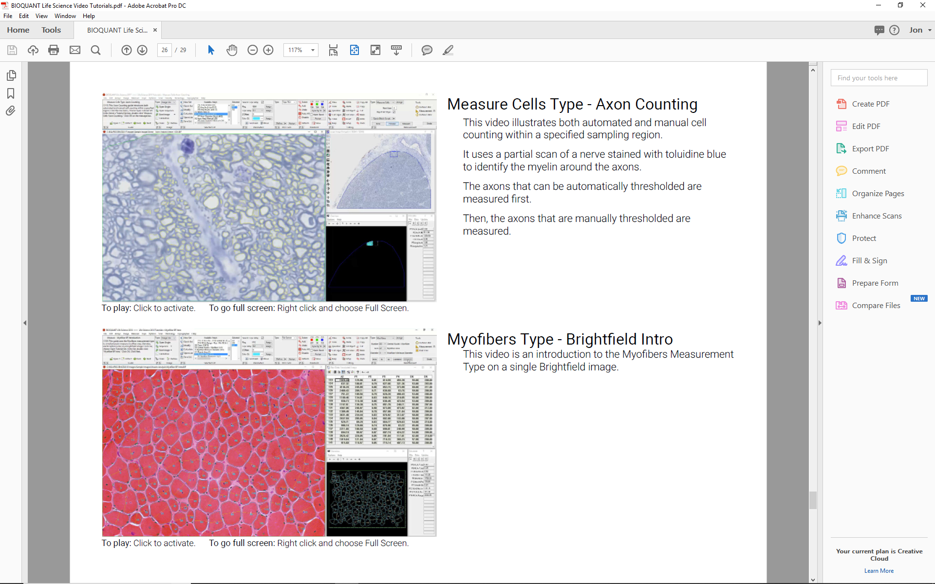
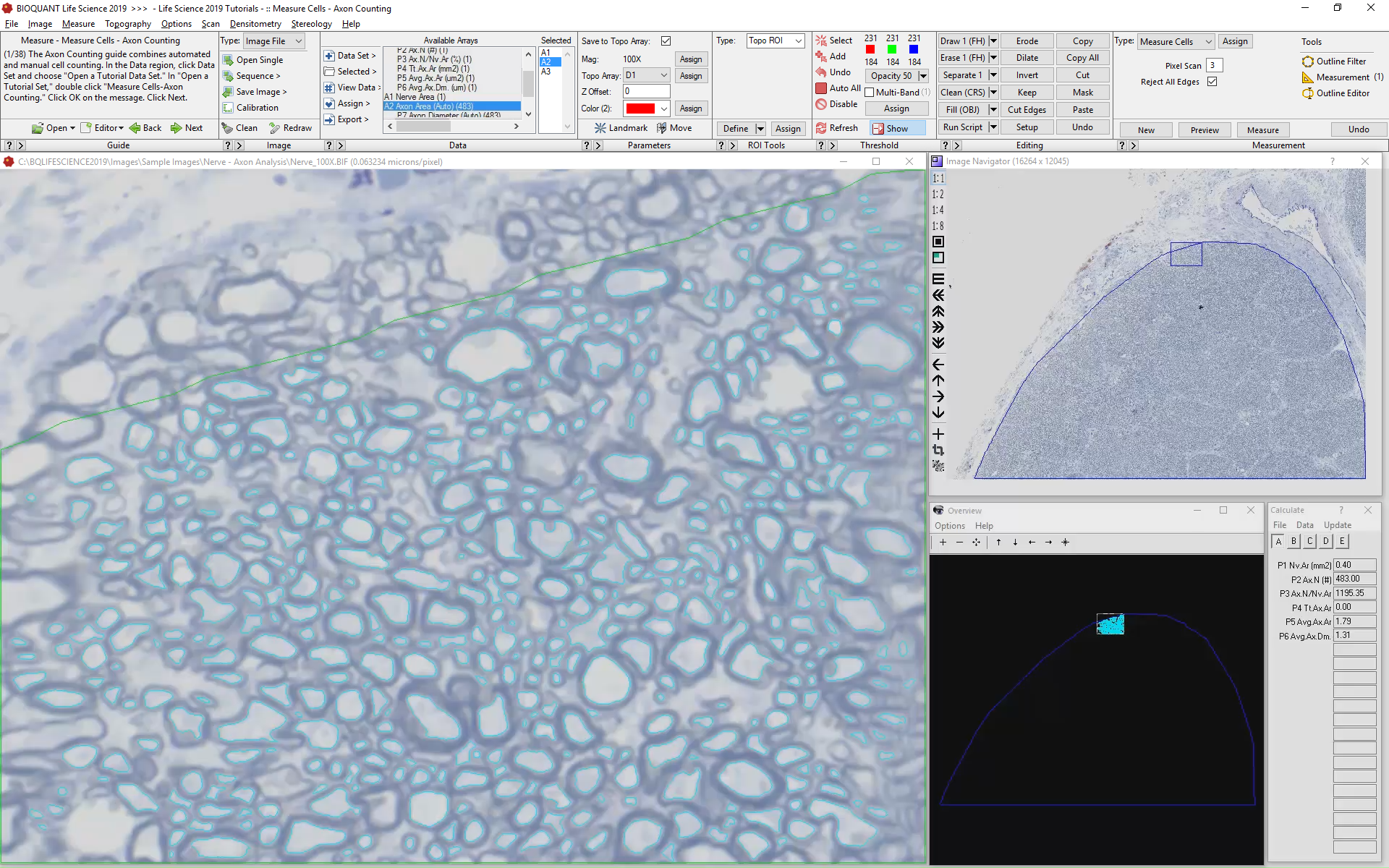
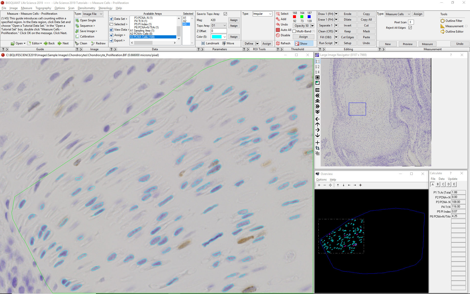
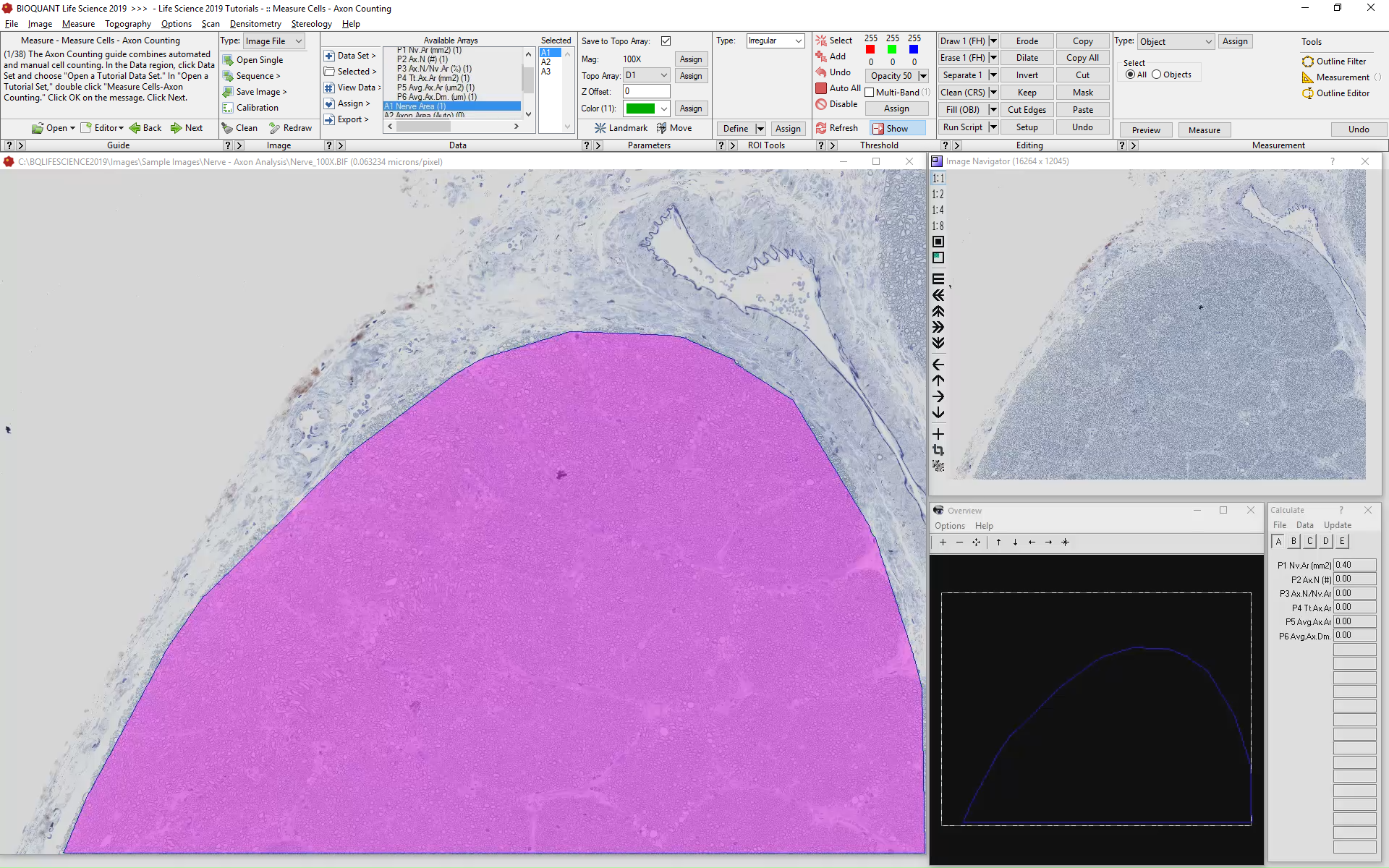
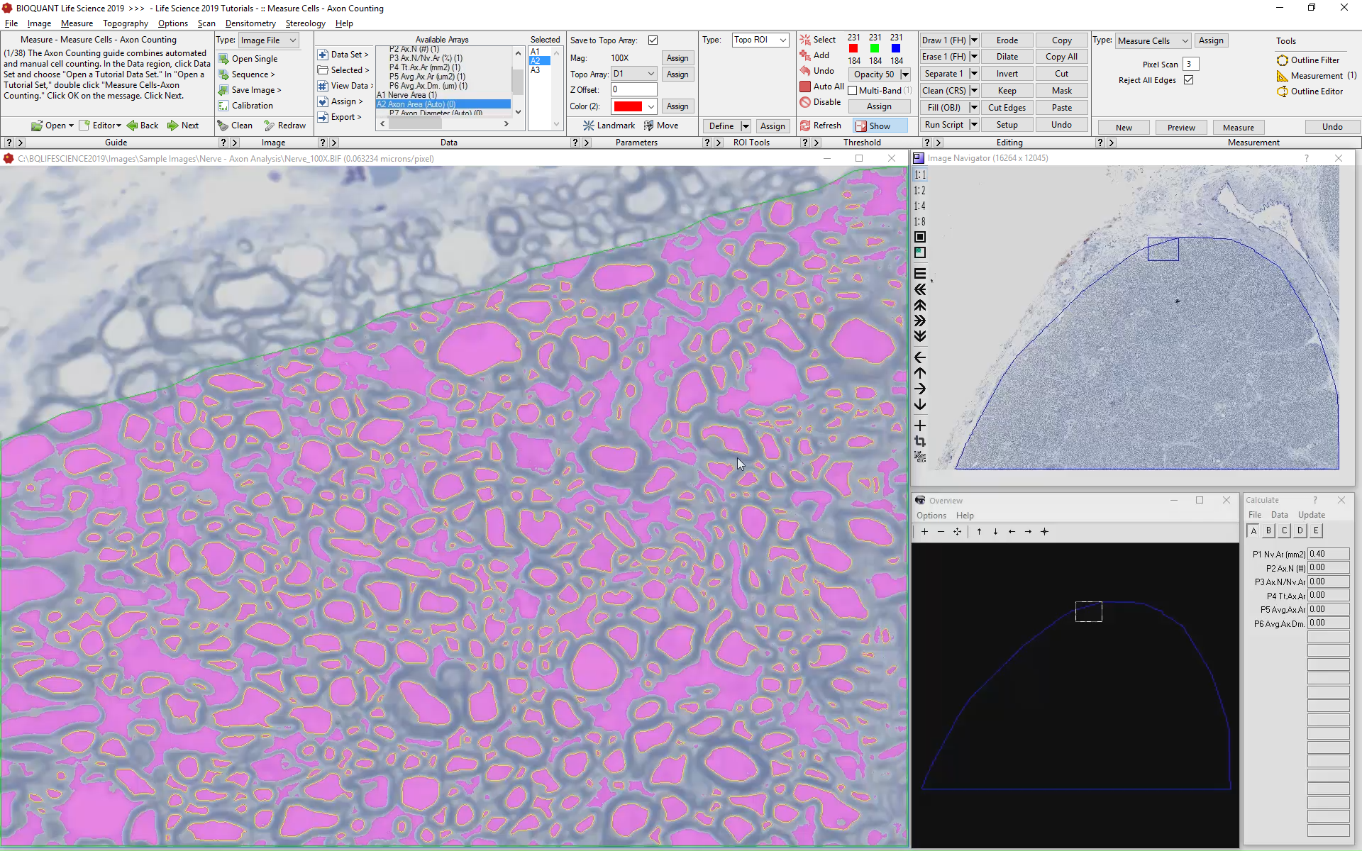
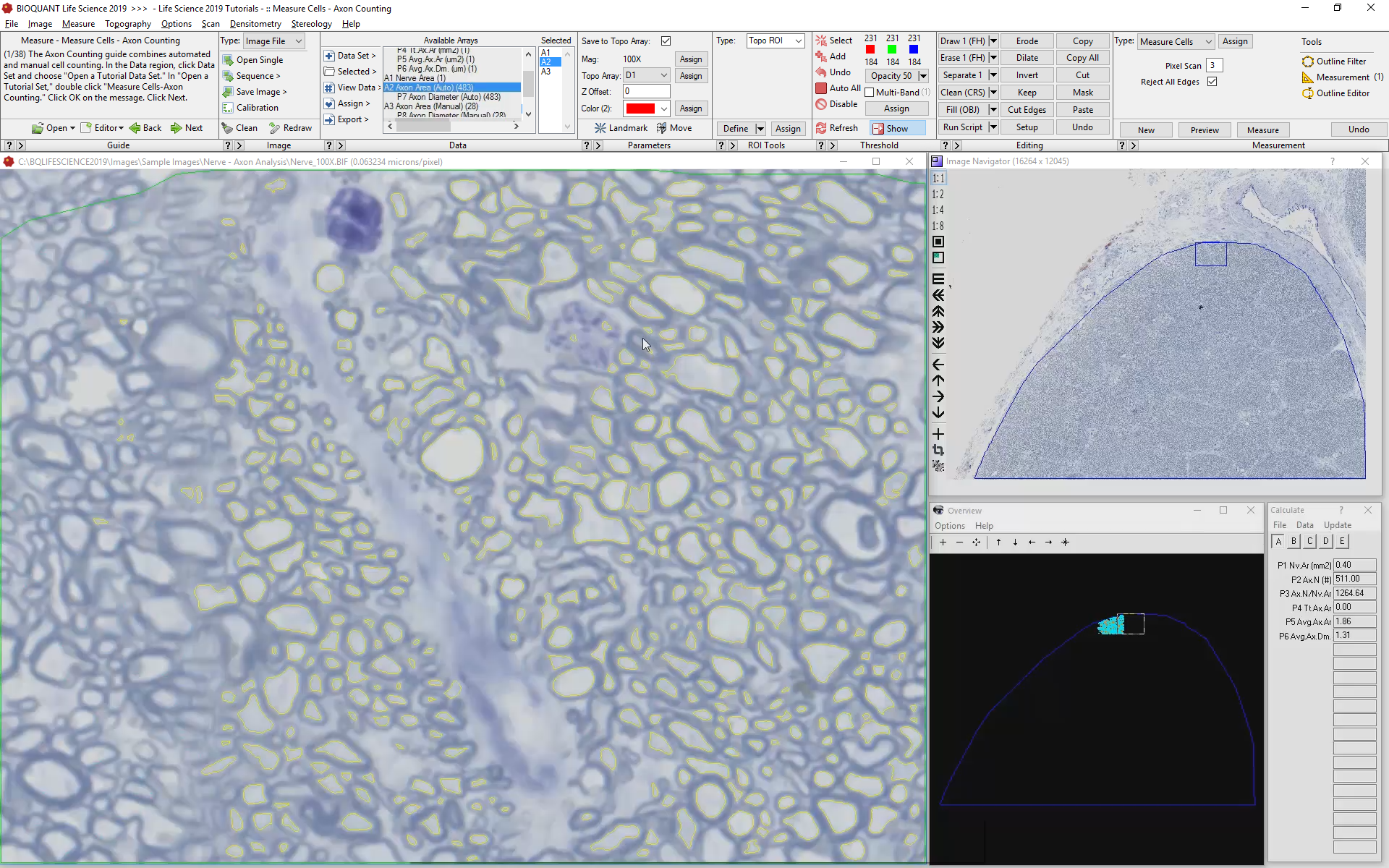
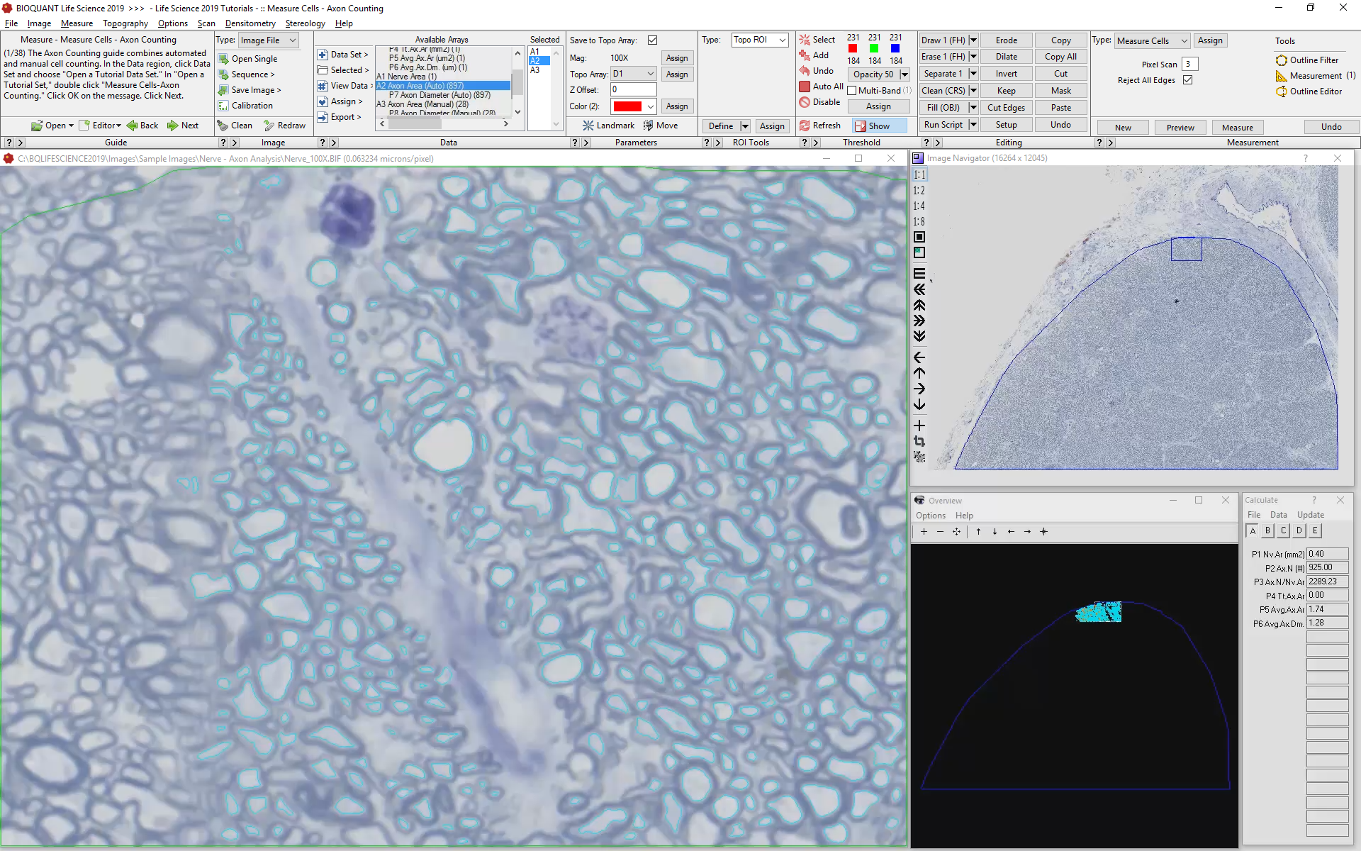
Automated Cell counting
One of the primary tasks of BIOQUANT is the automated analysis of cell populations. A specialized tool set makes this work fast and simple even for students who are new to image analysis. Here's an example of how the protocol works for the analysis of axon number, cross-sectional area, and diameter.

Automated analysis of Skeletal Myofibers
Analysis of skeletal muscle in particularly challenging because so many different parameters must be measured for each muscle cell. Most often these are: myofiber cross-sectional area, roundness, shortest diameter, position, optical density (for fibertyping), and number of central nuclei. BIOQUANT now includes a specialized tool precisely for this task.

rapid analysis of mri data
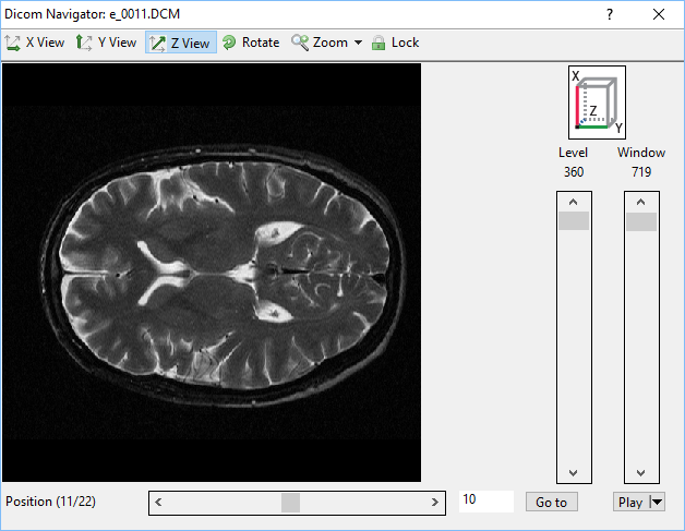
The new DICOM navigator for MRI data, quickly analyze the volume and shape of structures.
Compatible with batch processing, BIOQUANT can rapidly analyze stacks of DICOM format images.
To aid in visualizing data, a set of 8 new pseudocolor palettes make monochromatic data more vibrant.

Reason #9: Faster Help with Technical Service Plans

INDIVIDUAL Training via REMOTE DESKTOP
Labs with active Technical Service Plans receive on-demand training via remote desktop sharing. Use this service to train new students, learn new software features, and design new data collection protocols.
Custom Analysis Template Design
Work with our technicians to develop customized analysis templates. Start with one of our standard templates and improve it to match your samples. Or, build your own unique protocols from the ground up.
Priority technical support
Get quick solutions to software and hardware problems by phone, email, or remote desktop.
Continuous Software upgrades
Each year, BIOQUANT publishes an upgrade to BIOQUANT Life Science that is included at no charge as part of the Technical Services Plan. New versions add features requested by the community, consolidate protocol steps, and introduce new types of measurement tools.
Reason #10: Simply Faster
This version of BIOQUANT LIFE SCIENCE takes the longest strides toward distilling the complexity of analyzing biological tissues down to the simplest of procedures.
With a standardized workflow documented with both written and video tutorials, BIOQUANT combines intelligent object detection with simple tools for manual correction to rapidly analyze even the most complex biological samples. Let us help your lab today.